Reading time: 7 minutes
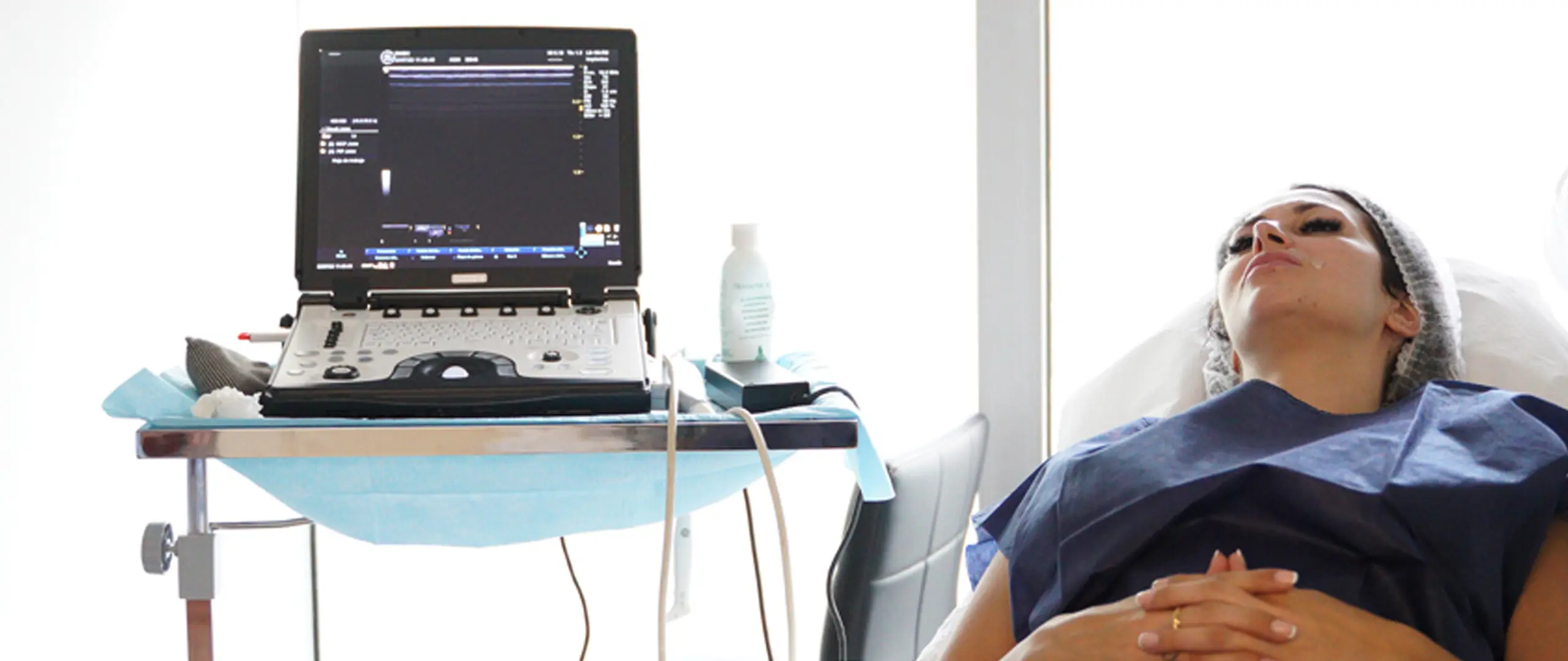
It gives light under the skin, allows a detailed observation and a comprehensive examination of the facial structure. But there is more, because aesthetic ultrasound is a diagnostic technique and a tool that offers precision, safety in each of the aesthetic treatments and the advantage of making an assessment not only of the epidermis, dermis, subcutaneous cellular tissue and superficial fat, but also of the musculature, deep fatty tissue and bone.
“Itallows us to have the best guarantee of where we are going to place the product, to assess each of the patients’ skin structures and, in this way, to avoid any type of vascular complication that may arise,” says Dr Oscar Jose Ramirez Prado, a specialist in family medicine and primary care, master in aesthetic medicine from the University of the Balearic Islands and fellow in dermo-aesthetic ultrasound.

Dr. Prado in a dermo-aesthetic ultrasound training.
In the vanguard
With the incorporation of this technique into the practice, it is also possible to determine what is currently known as the aesthetic footprint, something Prado says is key to understanding the history of each individual patient and, based on that, to indicate the best treatment. Spain has always been at the forefront, because this technique has also allowed us to assess the aesthetic footprint, in other words, previous treatments, fillers that the person may have had many years ago, such as biopolymers, silicone oil, polymethylmethacrylate; all those types of fillers that were used many years ago and which, when new treatments are carried out, always end up being a risk,” says the expert. The use of biopolymers, for example, which are not biocompatible, that is, they are not absorbable by the body. And this has generated a weight effect on the deep structures of the face and has also led to numerous complications in the superficial structures of the dermis. For this reason, and at the slightest doubt, it is advisable to have an ultrasound scan beforehand“.
Ultrasound, Prado adds, is not only useful at the time of diagnosis, prior to future treatment. There is also a trend towards the simultaneous use of ultrasound and the injection of facial fillers, known as ultrasound-guided protocols. “These are those in which we use ultrasound vision at the same time as we are assessing the placement of the facial filler. But we must differentiate between ultrasound-guided and ultrasound-guided treatments. The former are those in which vascular mapping is done at the puncture sites. An example: I want to place botulinum toxin at the level of the upper third, and I want to avoid puncturing the supratrochlear arteries [one of the terminal branches of the ophthalmic artery], which are those that can lead to blindness in the patient as a complication,” explains Prado. So, we mark the points on the patient where we are considering giving him his botulinum toxin treatment, and then we use ultrasound to assess with vascular mapping, i.e. where the arteries are, both in their position and in their plane. It can be in a superficial plane or in a deep plane. In this way, I avoid having any kind of complication. That is echodirected.

As for ultrasound-guided procedures, Prado explains with another example: “When I see a complication with hyaluronic acid deposits that can generate some kind of asymmetry, or some kind of vascular compression or decreased lymphatic drainage, generating water bags at the level of dark circles, for example. Using ultrasound, we can see where the hyaluronic acid deposit is, we can perforate with the syringe and place the enzyme that degrades hyaluronic acid, called hyaluronidase, directly on the affected tissue. This allows me to have safety and precision in each of the treatments,” emphasises the expert.
In addition to its advantages in non-surgical procedures, such as dermal fillers or the application of botulinum toxin, Prado mentions the usefulness of ultrasound for plastic surgery, such as liposuction. “Case one: the surgeon consults to perform an ultrasound scan to look at the patient’s abdominal wall and rule out hernias. Two: to assess the metastomeres; we all have the chocolates, we call them at the abdominal level, but what separates each of those chocolates at the level of the rectus abdominis are the metastomeres,” he explains. You can mark them for the plastic surgeon by saying ‘pass the cannula through this side to generate a high definition liposuction’. We can also do postoperative follow-ups to, for example, assess seromas or complications that arise after surgery. Ultrasound-guided and ultrasound-guided treatments to drain this type of seroma that may be generated by inflammatory or infectious processes after surgery,” concludes Prado, who has 16 years of experience in emergency medicine and critical patients.